Neurons Under the Microscope

Showing Commencement Weekend and throughout the summer 
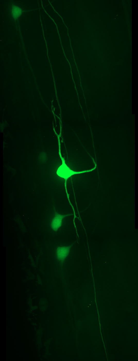
As you walk by Cabell Library, you may notice striking neon images that resemble abstract art. These are actually images of zebrafish spinal cord neurons, created as part of a master’s thesis in neurobiology by Ashley Purdy.
Purdy’s research studied the development of the nervous system and the mechanisms that guide neurons to their targets enabling the proper function of neural circuits. She focused on the multiple proteins that are necessary for cells to travel to their correct destination. Purdy disabled these proteins and examined the effect they had on the development of the nervous system. Her research builds towards a better understanding of neural development, which could assist in the comprehension and treatment of spinal cord injuries.
The transparent bodies of zebrafish allow Purdy to shine a laser onto the developing embryo to detect any changes. The use of zebrafish enables scientists to study this process in a live organism rather than in a cell culture. The images on the Cabell Screen are the products of these examinations.
Purdy used a Carl Zeiss spinning disc confocal microscope to create these images, and Zen Blue software to allow for maximum intensity projections. Her work was conducted in the laboratory of Dr. Gregory Walsh in VCU’s Department of Biology.
(This exhibition first ran in the spring semester, through April 14.)
Categories Science, Students